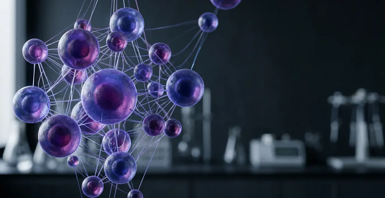
Mastering cellular interactions is no longer just an academic exercise; it is the core competency for a successful cancer research career within the unique UK ecosystem.
- Scientific breakthroughs are now driven by manipulating cellular dialogues, from immunotherapy to targeted drug delivery.
- The UK’s structure, blending NHS, charities, and industry, creates specific challenges and opportunities for translating this science into patient benefit.
Recommendation: View your scientific training not merely as knowledge acquisition, but as a strategic blueprint for navigating a career path from the lab bench to the patient’s bedside.
For any young biomedical scientist entering the field of cancer research, the complexity of cellular interactions can feel overwhelming. We are taught about the intricate dance of signalling pathways, the cascades of phosphorylation, and the delicate balance between cell life and death. The textbooks present this as a universe of molecular facts to be memorised. But this perspective misses the most crucial point, especially for those of us working within the United Kingdom’s unique scientific landscape. The common advice is to specialise, to pick one pathway and become the world’s expert on it. This is no longer enough.
The real challenge, and indeed the source of our greatest hope, lies not just in knowing these pathways but in understanding how to manipulate them within a living system—and, critically, how to navigate the ecosystem that turns a laboratory discovery into a clinical reality. In the UK, this means working at the intersection of world-class academic institutions, a publicly funded National Health Service (NHS), and a vibrant network of research charities. Understanding cellular interactions is not just about passing exams; it is about learning the language of the cells to develop therapies that can meet the rigorous efficacy and cost-effectiveness demands of bodies like NICE.
This is where the paradigm shifts. The true key is to see this deep scientific knowledge as a strategic asset. It’s the foundation upon which a meaningful career is built, a career that can genuinely contribute to medical breakthroughs. This isn’t about memorising diagrams; it’s about developing a mindset that connects a molecular mechanism to a patient outcome, a lab result to a funding application, and a scientific niche to a long-term career trajectory.
This article will guide you through that new perspective. We will explore the core scientific principles driving modern oncology and, crucially, connect them to the practical realities and career decisions you will face as a researcher in the UK. We will move from the science of cellular manipulation to the strategy of career navigation, showing how the two are inextricably linked.
Contents: A Strategic Guide to Cellular Interactions in UK Cancer Research
- How Checkpoint Inhibitors Block Negative Cellular Interactions?
- How to Visualize Cell Signaling Pathways in Real-Time?
- Apoptosis vs Necrosis: Why the Distinction Matters for Drug Toxicity?
- The Incubator Error That Ruins Weeks of Cellular Interaction Data
- How to Target Specific Cellular Receptors to Reduce Chemo Side Effects?
- The Specialist Trap: Why Staying in One Niche Too Long Hurts Mobility
- How to Find the Secret Job Boards Used by Charity and NGO Sectors?
- NHS vs Private Sector: Where to Start Your Biomedical Engineering Career?
How Checkpoint Inhibitors Block Negative Cellular Interactions?
The advent of immune checkpoint inhibitors represents one of the most significant paradigm shifts in cancer therapy, and it is a story rooted entirely in manipulating cellular interactions. In essence, many cancers have learned to exploit natural “off-switches” on immune cells, primarily T-cells, to evade destruction. They present proteins like PD-L1 on their surface, which bind to the PD-1 receptor on an activated T-cell. This interaction is a negative signal; it tells the T-cell to stand down, effectively cloaking the tumour from the immune system.
Checkpoint inhibitors are monoclonal antibodies that physically block this conversation. An anti-PD-1 drug, for instance, will bind to the PD-1 receptor on the T-cell, preventing the cancer cell’s PD-L1 from docking. This action removes the “brake” and allows the T-cell to recognise and attack the cancer cell. It’s a beautifully simple concept: we are not killing the cancer directly, but rather enabling our own immune system to do its job by blocking a single, negative cellular interaction. The scale of this research in the UK is vast; leading centres like the Royal Marsden’s Drug Development Unit are conducting over 75 early phase trials annually, giving hundreds of patients access to these next-generation treatments.
Case Study: The UK Cancer Drugs Fund and Managed Access
Getting these promising but expensive drugs to patients presents a unique challenge in the UK. The reformed Cancer Drugs Fund provides a powerful solution through a ‘managed access’ pathway. This system, a partnership between NHS England and NICE, allows promising checkpoint inhibitors to be used in the NHS for a period of up to two years while more real-world data on their effectiveness and cost-efficiency is gathered. It’s a pragmatic British approach that balances the urgent need for innovation with the fiscal responsibilities of the NHS, demonstrating how understanding the national healthcare ecosystem is as crucial as understanding the science.
For a junior researcher, this field isn’t just about understanding the PD-1 pathway. It’s about grasping how biomarkers can predict which patients will respond, and how resistance mechanisms—new negative interactions—evolve. This is the frontier of immuno-oncology.
How to Visualize Cell Signaling Pathways in Real-Time?
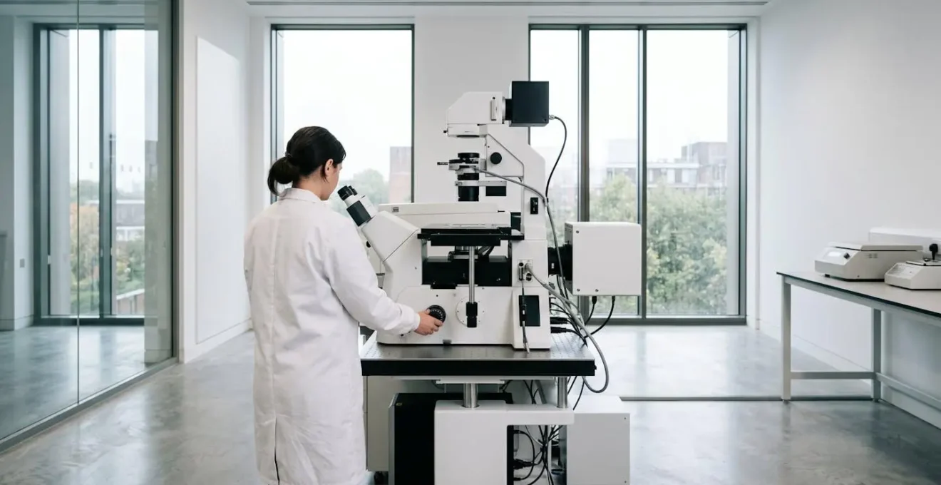
Understanding cellular conversations is one thing; seeing them happen is another. The ability to visualize these dynamic processes in real-time is what separates theoretical knowledge from actionable discovery. Modern cancer research labs in the UK are powered by advanced imaging technologies that turn the abstract concept of a signalling cascade into a vivid, measurable event. Techniques like confocal microscopy, two-photon microscopy, and FRET (Förster resonance energy transfer) allow us to watch proteins interact, ions flow, and cells respond to stimuli with incredible precision.
These are not just pretty pictures. By tagging proteins with fluorescent markers, we can quantify the activation of a pathway. We can see if a drug is hitting its intended receptor, or if it’s causing unintended off-target effects in healthy cells. This visual data is the bedrock of preclinical drug development. The UK’s commitment to this field is substantial; a recent UKRI announcement detailed a £473 million total infrastructure investment, which includes massive funding for next-generation imaging facilities. This investment ensures that UK-based researchers have the tools to remain at the cutting edge.
As you can see, these facilities are the cathedrals of modern biology, where light is manipulated to reveal life’s most fundamental secrets. Working in such an environment requires a hybrid skillset—a deep understanding of cell biology combined with a technical grasp of optics, software, and data analysis. As Dr. Diana Passaro, a leading scientist at the Francis Crick Institute, explains, the goal is always translation:
We’re starting by imaging mice, but if our findings are promising, it could have direct implications for diagnostic and prognostic approaches in human patients.
– Dr. Diana Passaro, Francis Crick Institute, Medical Research Foundation feature on Crick imaging progress
This captures the spirit of pragmatic hope that drives our work. The images we capture today are the diagnostics of tomorrow.
Apoptosis vs Necrosis: Why the Distinction Matters for Drug Toxicity?
At the heart of many cancer therapies is a simple goal: induce cell death in malignant cells while sparing healthy ones. However, the way a cell dies is critically important. For decades, the focus was on apoptosis, or programmed cell death. This is an orderly, controlled process where the cell dismantles itself from within, packaging its contents into neat bundles that are cleaned up by immune cells. It’s a “clean” death that doesn’t provoke an inflammatory response. In contrast, necrosis is a messy, chaotic death, often caused by acute injury. The cell swells and bursts, spilling its contents into the surrounding tissue and triggering a strong inflammatory cascade. This inflammation can be highly damaging and is a major source of drug toxicity and side effects.
For a drug developer, designing a compound that selectively induces apoptosis in cancer cells is the holy grail. A drug that causes widespread necrosis, on the other hand, is likely to be too toxic for human use. This distinction is a key reason why so many promising drugs fail in clinical trials. A therapy might kill cancer cells effectively in a petri dish, but if it does so by causing necrosis in vital organs, it will never be approved. In fact, a sobering analysis of drugs in the pre-2016 Cancer Drugs Fund showed how few met criteria for meaningful clinical benefit, often due to a poor balance between efficacy and toxicity.
Today, our understanding is even more nuanced, as a leading UK expert highlights:
Our improved understanding of the different types of cell death – apoptosis, necroptosis, pyroptosis, and ferroptosis – and their interconnectivity with innate and adaptive immunity has unearthed new opportunities to guide our immune system to fight cancer.
– Professor Pascal Meier, Institute of Cancer Research Cell Death and Immunity Programme
This growing knowledge of different cell death modalities—some inflammatory, some not—is opening up new therapeutic avenues. We can now design drugs that not only kill cancer but do so in a way that stimulates a helpful immune response, turning a potential liability (inflammation) into a therapeutic asset.
The Incubator Error That Ruins Weeks of Cellular Interaction Data
All the advanced science and brilliant theory we’ve discussed can be rendered meaningless by a single, mundane failure in the lab. Perhaps the most devastating—and common—of these is the incubator error. A cell culture incubator is a precisely controlled environment, maintaining a constant 37°C, high humidity, and a 5% CO2 atmosphere to keep the culture medium’s pH stable. It is the sterile womb where our precious cells, the subjects of our experiments, live and grow. A failure here is catastrophic.
Imagine this scenario: you have been treating a co-culture of cancer cells and immune cells with a novel compound for two weeks. Your experiment is designed to measure the subtle changes in cytokine secretion and T-cell activation. On Monday morning, you arrive to find the incubator’s CO2 tank is empty. The alarm, for whatever reason, failed to alert anyone over the weekend. The pH of your culture medium has skyrocketed, becoming alkaline. Your cells are dead or dying, not from your drug, but from an environmental shock. Two weeks of work, the cost of the reagents, the use of the expensive cell lines—all of it is gone. Worse, if the failure was subtle (e.g., a slight temperature drift or a low-level contamination), the data might not be obviously wrong, leading you to draw false conclusions that could waste months of follow-up work.
This is not a hypothetical. It is a rite of passage for many a PhD student and a constant source of anxiety for a Principal Investigator. It underscores a vital truth: rigour in basic lab practice is the non-negotiable foundation of good science. Without it, the most sophisticated understanding of cellular interactions is worthless.
Your Lab-Based Pre-Flight Checklist: Preventing Data Catastrophe
- Daily System Checks: Physically check CO2 tank pressure, water pan levels, and temperature display every morning. Do not rely solely on digital monitoring.
- Redundant Alarms: Ensure your incubator is connected to a remote alarm system that can text or call multiple lab members in case of a failure outside of working hours.
- Routine Culture Screening: Regularly test a sample of your cultures for mycoplasma contamination, the invisible data-killer. Schedule this for the first Friday of every month.
- Sensor Calibration: Implement a bi-annual professional calibration schedule for the incubator’s CO2 and temperature sensors. A drifting sensor gives a false sense of security.
- Data Logging: Use an independent data logger inside the incubator to record temperature and CO2. This provides an objective record to validate experimental conditions or diagnose a subtle problem.
How to Target Specific Cellular Receptors to Reduce Chemo Side Effects?
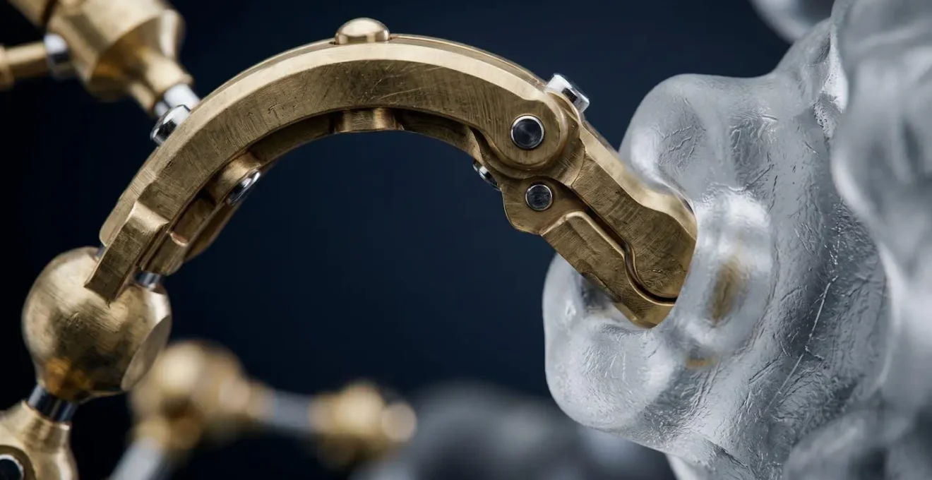
For decades, the foundation of chemotherapy was cytotoxic drugs that kill any rapidly dividing cell. This is why they are effective against cancer, but also why they cause such severe side effects, damaging hair follicles, the gut lining, and bone marrow. The future of oncology lies in precision: delivering a lethal payload only to cancer cells by targeting unique receptors on their surface. This is the world of Antibody-Drug Conjugates (ADCs), and it is a perfect marriage of immunology and pharmacology.
An ADC is a three-part molecule: a monoclonal antibody, a highly potent cytotoxic drug, and a chemical linker. The antibody is engineered to bind with high specificity to a receptor that is overexpressed on cancer cells but largely absent from healthy cells (e.g., HER2 in some breast cancers). The ADC circulates harmlessly in the body until its antibody finds and binds to its target receptor on a cancer cell. This binding triggers the cell to internalise the ADC. Once inside, the linker is cleaved by cellular enzymes, releasing the cytotoxic drug directly within the malignant cell, leading to its death. It’s like a biological smart bomb.
This lock-and-key mechanism dramatically reduces collateral damage to healthy tissues, allowing for the use of toxins that would be far too potent to be administered as standard chemotherapy. The success of this approach depends entirely on a deep understanding of the cellular landscape. Which receptors are truly unique to the cancer? How quickly are they internalised? What linker chemistry will be stable in the bloodstream but labile inside the cell? These are the questions that drive ADC development, a major focus for UK biotech and pharmaceutical companies.
For a young scientist, this field offers a chance to work at the cutting edge of protein engineering, linker chemistry, and cell biology. It is the ultimate expression of turning our knowledge of cellular interactions into a smarter, kinder form of medicine, moving beyond blunt instruments to highly specialised tools.
Key Takeaways
- Manipulating cellular interactions (e.g., with checkpoint inhibitors) is the basis of modern cancer immunotherapy.
- Advanced imaging, heavily funded in the UK, is essential for visualizing and validating these interactions in real-time.
- The distinction between different cell death pathways (apoptosis vs. necrosis) is critical for creating effective drugs with low toxicity.
The Specialist Trap: Why Staying in One Niche Too Long Hurts Mobility
The traditional academic path encourages deep specialisation. You do a PhD on the Wnt signalling pathway, a postdoc on a specific kinase within that pathway, and aim to become the world’s leading expert on Wnt-regulated kinase X. While expertise is valuable, in the rapidly evolving landscape of cancer research, hyper-specialisation can become a career trap. Science does not stand still. A pathway that is central to the field today might be superseded by a new discovery tomorrow. A class of drugs that seems revolutionary can be rendered obsolete by a new modality with fewer side effects.
If your entire skillset is tied to a single target or technique, you risk becoming a scientific dinosaur. When funding priorities shift or a new technology emerges, you may find your deep expertise is in a suddenly shrinking field. Mobility—the ability to adapt your skills to new questions and new areas of biology—is paramount for long-term career stability and impact. This doesn’t mean being a jack-of-all-trades. It means building a T-shaped skillset: a deep expertise in one core area (the vertical bar of the T) combined with a broad understanding of related fields, techniques, and concepts (the horizontal bar).
For example, if you are an expert in a specific signalling pathway, you should also have a working knowledge of bioinformatics to analyse transcriptomic data, an understanding of advanced microscopy to visualize your pathway’s activity, and an appreciation for the clinical challenges that your research might one day address. This breadth allows you to pivot. When a new pathway is discovered to interact with yours, you can lead the charge. When a new technology like CRISPR becomes available, you can apply it to your system. You are no longer just a “Wnt pathway person”; you are a cancer biologist who can solve problems using a diverse toolkit.
How to Find the Secret Job Boards Used by Charity and NGO Sectors?
When searching for a research position, many graduates default to the big, generic academic job portals. However, a huge portion of UK cancer research is funded and conducted by charities and non-governmental organisations (NGOs), and they often use more specialist channels. There are no “secret” job boards, but there are certainly more focused and efficient places to look. Ignoring these is a significant missed opportunity, as these roles often provide a unique blend of cutting-edge science and a clear, mission-driven purpose.
The first and most important platforms are the charity-specific job sites. Websites like CharityJob and Third Sector Jobs are the primary recruitment hubs for the UK’s non-profit world. Major research charities like Cancer Research UK (CRUK), the Wellcome Trust, and the British Heart Foundation will post everything from lab-based research assistant roles to policy and grant management positions on these sites, as well as on their own dedicated career portals.
Secondly, do not underestimate the power of university networks, but look beyond the central HR page. Technology Transfer Offices (TTOs) and university-linked “incubator” or “accelerator” hubs are fantastic places to find roles in spin-out companies that are commercialising academic research. Finally, targeted networking is key. Attending specific UK conferences, such as the NCRI Cancer Conference or smaller, topic-focused symposia hosted by institutes like the ICR or the Crick, puts you in direct contact with the PIs and hiring managers from these organisations. Following key labs and charities on professional networks like LinkedIn or even academic Twitter can also provide early leads on upcoming positions before they are widely advertised.
NHS vs Private Sector: Where to Start Your Biomedical Engineering Career?
For a biomedical scientist or engineer in the UK, a fundamental career crossroads is the choice between the public sector, primarily the NHS, and the private sector, which spans from nimble biotech start-ups to global pharmaceutical giants. There is no single “right” answer; the optimal choice depends entirely on your personal career goals, risk tolerance, and what you find most motivating. Understanding the distinct culture and opportunities of each is essential for making an informed decision.
A career within or closely aligned with the NHS offers unparalleled proximity to the patient. You might be working in a hospital lab, helping to implement new diagnostic tests, or in a university lab that collaborates directly with clinicians. The work has a tangible, immediate sense of impact. The career path is often more structured and stable, with clear progression routes and public sector benefits. However, the pace can be slower, constrained by bureaucracy and budget cycles. The focus is often on the implementation and optimization of existing technologies rather than blue-sky invention.
The private sector, in contrast, is driven by innovation and speed. In a biotech start-up, you could be working on a truly novel therapeutic modality, with the potential for high risk and high reward. The environment is dynamic, fast-paced, and you may have a greater degree of autonomy. In big pharma, you will have access to immense resources and be part of a global effort to bring new drugs to market. The trade-off can be a disconnect from the final patient, a higher-pressure environment, and less job security, particularly in smaller companies that are reliant on venture capital funding. Answering the question “Is a PhD necessary?” also differs; it’s often a prerequisite for leadership in R&D, but less so for applied roles in both sectors.
Ultimately, the UK’s strength lies in the porous border between these two worlds. Many successful careers involve moving between them, bringing commercial acumen to an academic project or clinical insight to a private enterprise. Seeing them not as a binary choice but as two essential parts of a single “bench-to-bedside” ecosystem is the key to a dynamic and impactful career.
Your deep understanding of cellular interactions is your passport to either world. The next step is to consciously build a career strategy that aligns your scientific passion with the environment where you can make your greatest contribution. Start by identifying the research groups and companies—in both sectors—that are asking the questions you find most exciting.